最高のコレクション t l spine x ray 168805-T l spine x ray
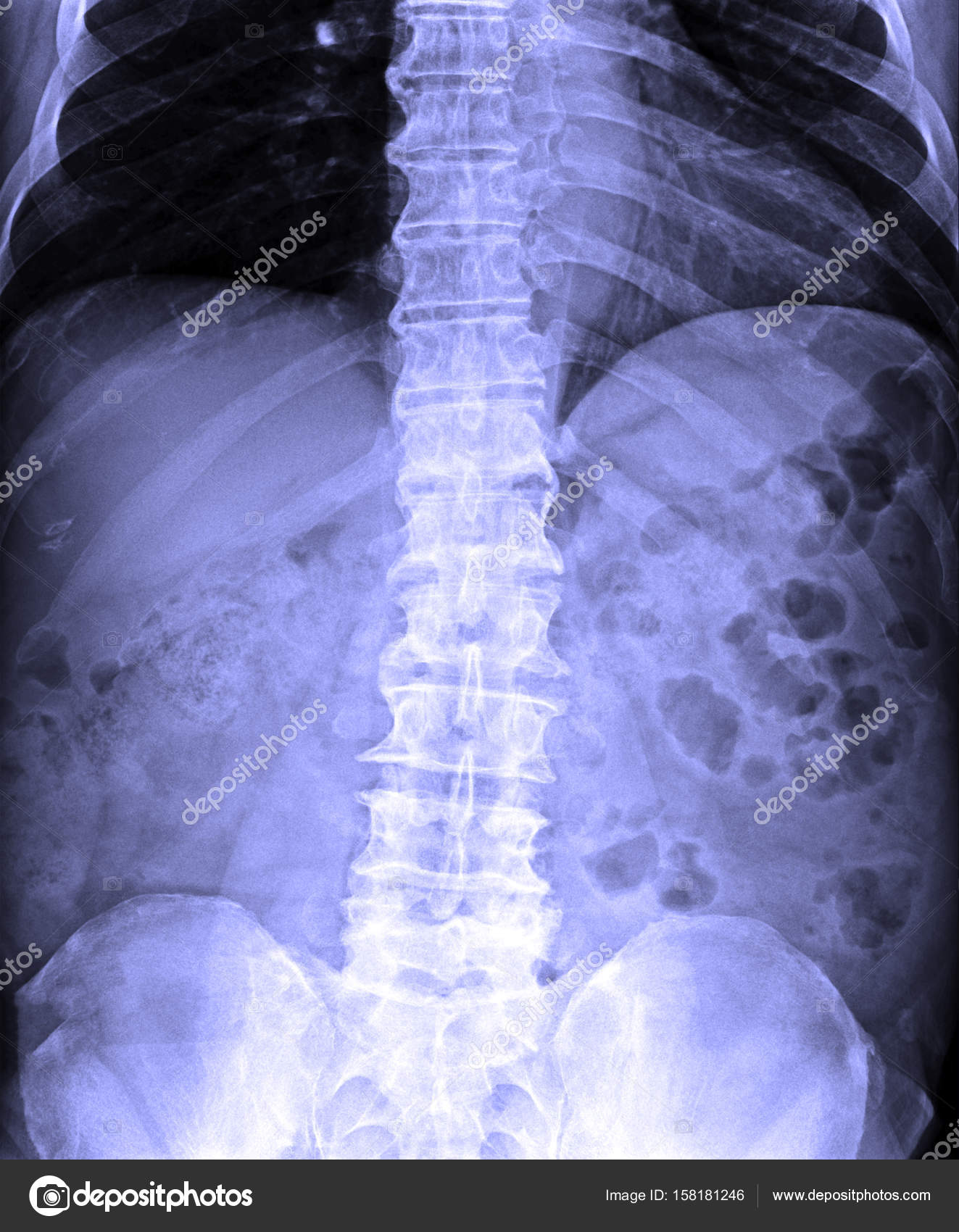
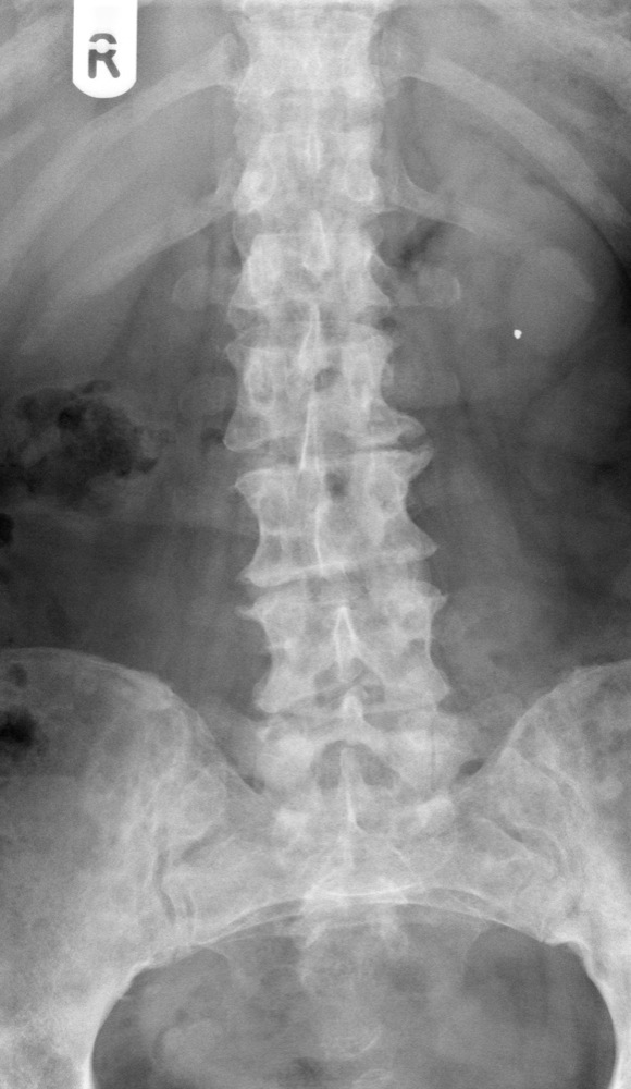
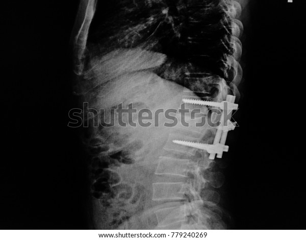
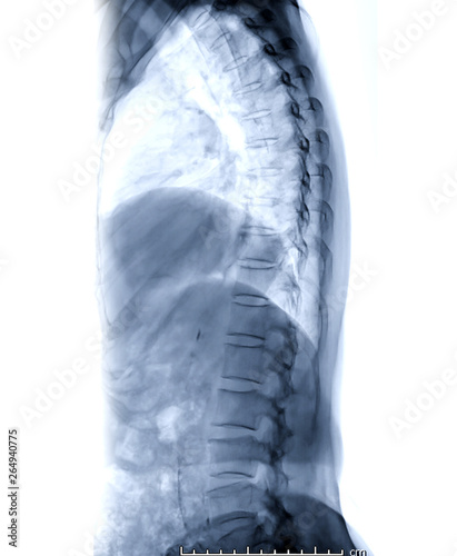
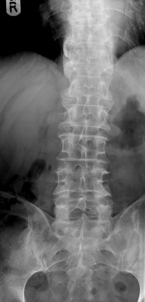
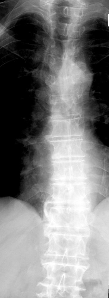
Film xray TL spine(ThoracicLumbar spine) show human's thoraciclumbar spine and inflammation at spine Film xray TL spine (ThoracicLumbar spine) show Film xray Skull lateral view show normal human& x27;s skull and cervical spine and blank area at left sideFor a lumbar spine view you should be able to see L1L5 but also the full T12 vertebral body, T11/12, and the sacrum on the AP viewA thoracic spine Xray is an imaging test used to inspect any problems with the bones in the middle of your back An Xray uses small amounts of radiation to see the organs, tissues, and bones of
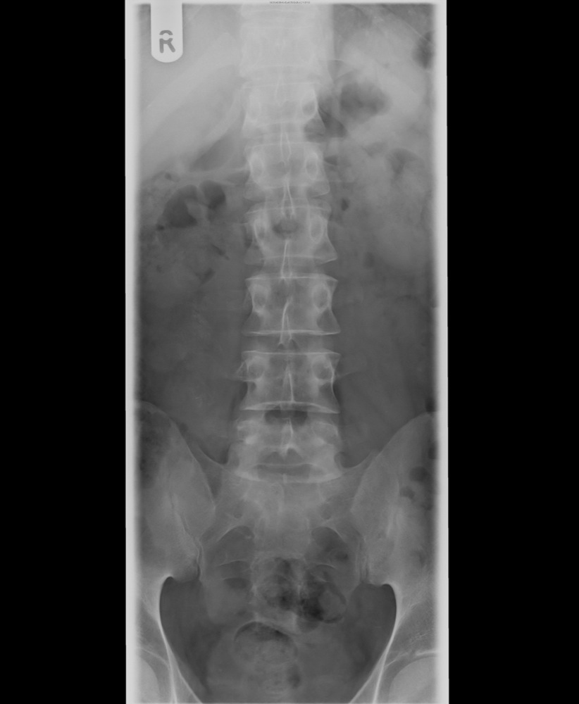
The Thoracolumbar Spine
T l spine x ray
T l spine x ray-ThoracoLumbar Spine Xray Guideline Routine 2 or 4 views • AP Thoracic spine • AP Lumbar spine • LATERAL Thoracic spine • LATERAL Lumbar spine • 2 VIEW is centered on junction of ThoracoLumbar spine • AP AND LATERAL VIEWS to include T8 thru L5 517 AMRFilm xray TL spine (ThoracicLumbar spine) show human's thoraciclumbar spine and inflammation at lumbar spine Man filling customer survey form on tablet computer Xray image pelvic bone and part of femur, spine Film xray of normal human lumbar spine
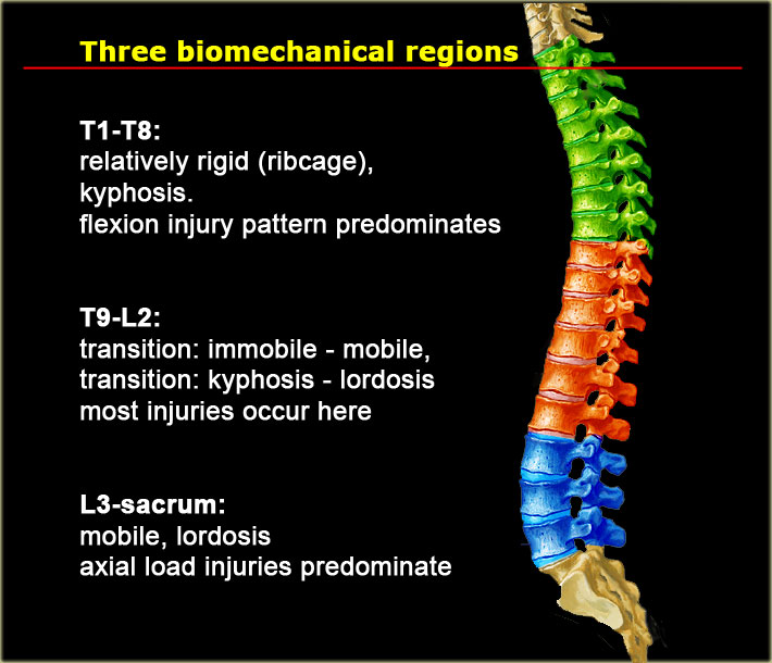


The Radiology Assistant Thoracolumbar Injury
RayT6(xiphoid process 위쪽3cm),jugular notch아래8~10cm호흡은 full exhalation후 멈춘다음에 x선조사를 한다collimation 의 위 margin을 턱선에 맞춰서 검사한다12개의 흉추(thoracic spine)가 모두포함되어야한다thoracic spine 가급적 균일한 음영으로 보여야한다 Tspine Lateral 촬영목적XRay Report Cervical Spine AP and lateral cervical spine views provided Complete loss of the normal cervical lordosis with mild reversal centred at C5/6 measuring 41° with 25mm anterior head carriage A 5° left lateral list extends from the lower cervical spine with left inferior occiputA lumbosacral spine Xray, or lumbar spine Xray, is an imaging test that helps your doctor view the anatomy of your lower back The lumbar spine is made up of five vertebral bones The sacrum is
A lateral spine Xray is when an Xray is taken from the side so a radiologist can evaluate your vertebrae This test is noninvasive and will not hurt You will usually lie on a table so the radiology tech is able to get a good image The Xray is very quick and you will only need to stay still for a short amount of time A lateral spine Xray is recommended every 12 yearsA, An adequate lateral cervical spine xray, showing the entire cervical spine and its cephalad border (the skull base) and caudad border (T1) B, The image is inadequate because C1 and T1 are not seen fully Fractures or subluxation at these locations would be missedA spinal Xray is a procedure that uses radiation to make detailed pictures of the bones of your spine It can help your doctor find out what's causing your back or neck pain A technician uses a
Explanation of the anatomy of the lumbar spine on xrayA Thoracic Spine XRay may help diagnose (find) Thoracic spine Xrays can detect fractures in the thoracic vertebrae or dislocation of the joints between the vertebrae Xrays of the thoracic spine can also find the cause of tingling, numbness, or weakness in the arm or hand A physician typically requests an Xray of the thoracic spine after a severe accident resulting in an injury to the head, neck or backThoracic Spine AP or PA Oblique Projection Upright Purpose and Structures Shown An oblique projection of the thoracic spine to demonstrate the zygapophyseal articulations Position of patient Standing or sitting erect in a lateral position in front of a vertical gridThe body is rotated degrees posterior or anterior (AP or PA oblique, respectively)


Adult Spinal Deformity The Lancet


Film X Ray T L Spine Thoracic Lumbar Spine Show Human Gl Stock Images
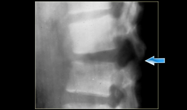
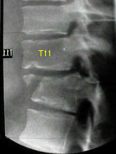
Edge of image In the context of trauma similar principles apply to imaging both the Thoracic spine (Tspine) and the Lumbar spine (Lspine) The plain Xray anatomy and appearances of injuries to both these areas are discussed together Incorrect management of patients with spinal injury may cause or worsen neurological deficitFilm xray TL spine (ThoracicLumbar spine) show human s thoraciclumbar spine and inflammation at spineRadiographs of the thoracic spine are considered the basic primary imaging, having a far inferior diagnostic yield than that of CT and MRI 1 Indications Thoracic spine radiographs are performed for a variety of indications including 1,2 fall from a height of greater than 3 meters;


Thoracolumbar Spine Fracture Radiology Reference Article Radiopaedia Org


The Radiology Assistant Thoracolumbar Injury
Dynamic Xrays of the Lumbar Spine How Useful Are They?Dynamic Xrays of the Lumbar Spine How Useful Are They?Standing AP and lateral Xrays are often ordered when back and/or leg pain doesn't go away The value of Xrays in diagnosing low back pain (LBP) has been questioned in the past In this study, researchers reviewed the use of flexionextension (F/E) Xrays for patients with LBP
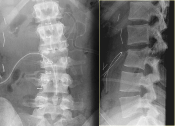


The Radiology Assistant Thoracolumbar Injury


Small Animal Spinal Radiography Series Thoracic Spine Radiography Today S Veterinary Practice
A thoracic spine xray is an xray of the 12 chest (thoracic) bones (vertebrae) of the spine The vertebrae are separated by flat pads of cartilage called disks that provide a cushion between the bones How the Test is Performed The test is done in a hospital radiology department or in the health care provider's officePhoto about Xray image of TL spine, AP view Showing a compression fracture at T12 Image of bone, joint, careLumbosacral Spine XRay (XRay) This is another useful X Ray which helps doctor view the anatomy of your lower back The lumbar spine is basically made up of five vertebral bones besides blood vessels, nerves, tendons, cartilage and ligaments etc The test is ordered in case of pain in lower back due to accident or fall


Thoracolumbar Spine X Rays


X Ray T L Spine Stock Photo Picture And Royalty Free Image Image
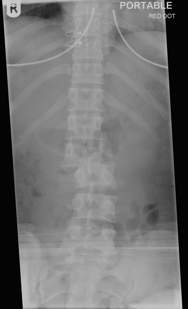
Lumbosacral Spine XRay (XRay) This is another useful X Ray which helps doctor view the anatomy of your lower back The lumbar spine is basically made up of five vertebral bones besides blood vessels, nerves, tendons, cartilage and ligaments etc The test is ordered in case of pain in lower back due to accident or fallRead our stepbystep guide to interpreting thoracic and lumbar spine xrays Thoracolumbar spine xray involves two views – AP and lateral Check it's an adequate view;In order to replicate the conditions under which there is too much movement in the spine vertebrae, an xray can be taken when the patient moves This is called a flexionextension xray For the low back, the patient is asked to bend forward and then backwards while xray images are taken in both positions


Q Tbn And9gcssrd2uhsflsle2mkqkm84bgfa32vsd Ugyq5btoxaoeccapm Usqp Cau
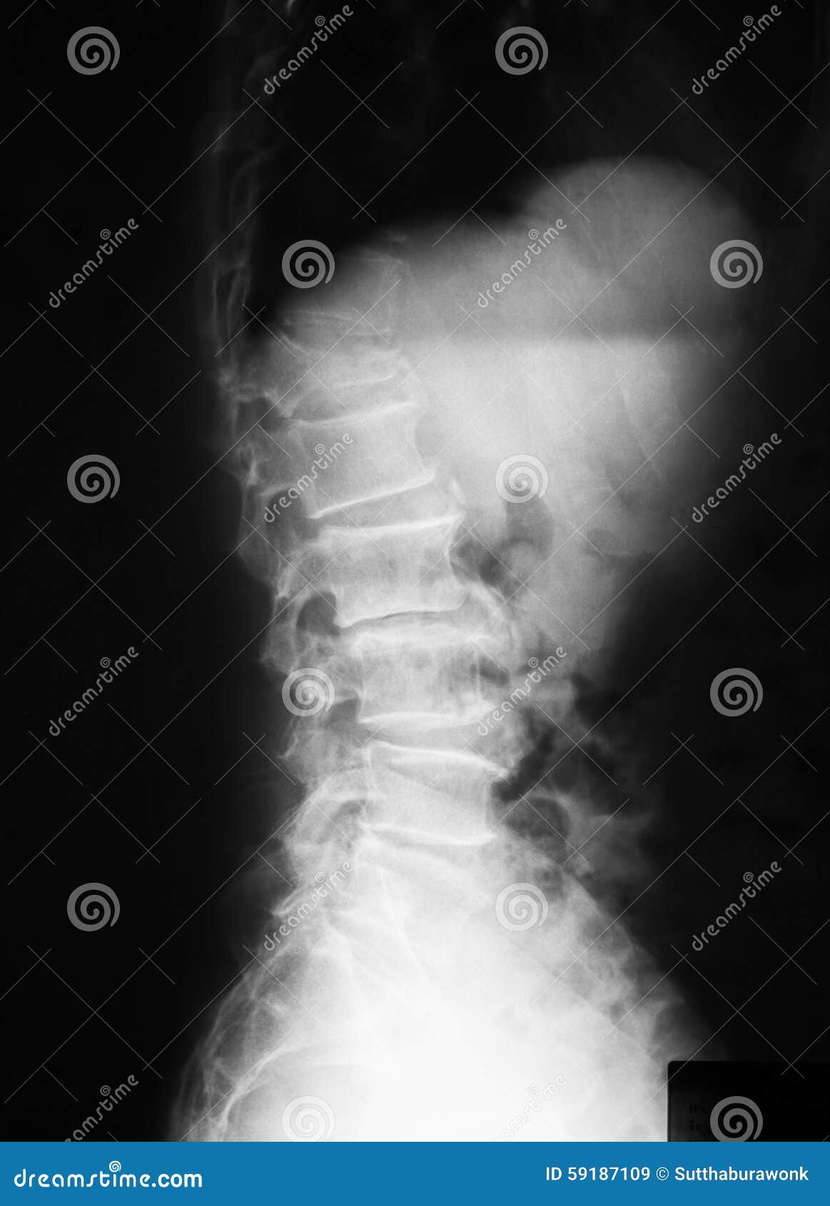


X Ray Image Of T L Spine Lateral View Stock Image Image Of Compression Lumbar
The thoracic spine xray is the imaging of the twelve bones extending from upper and middle back portion of our neck The thoracic spine joins the cervical spine and further connects with lumbar spine The Xray can spot for injuries in the thoracic spine In a human body a spine is made up of three subsectionsThe lumbar spine anteroposterior or posteroanterior view images the lumbar spine in its anatomical position The lumbar spine generally consists of five vertebrae (see lumbosacral transitional vertebra)A CT scan of the thoracic spine images the 12 vertebrae in the middle portion of the spine Your thoracic spine spans from the base of the neck to the middle of your back, running the length of your chest This exam is also called a Tspine CT
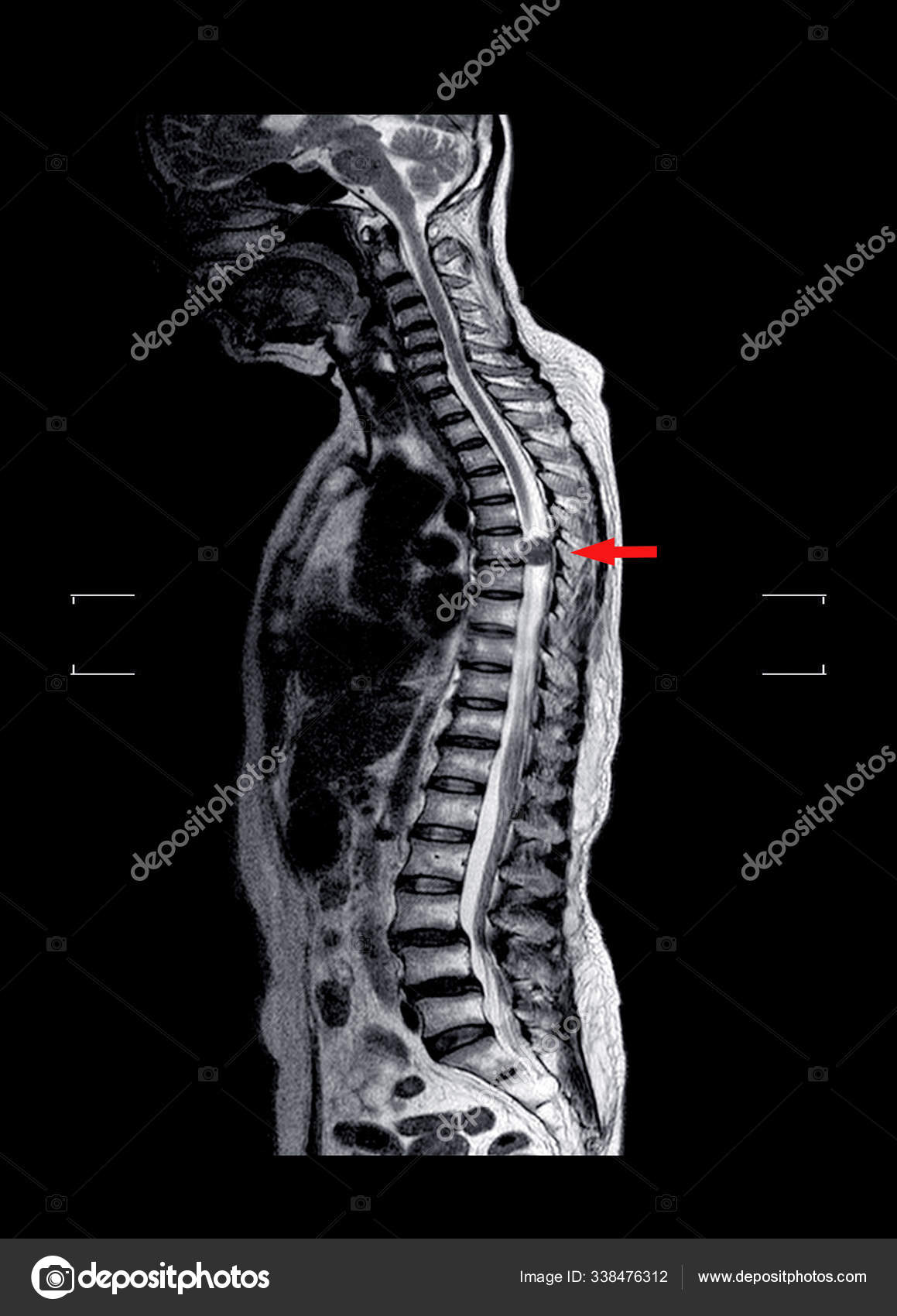


Mri Tl Spine Stock Photo Image By C Richmanphoto
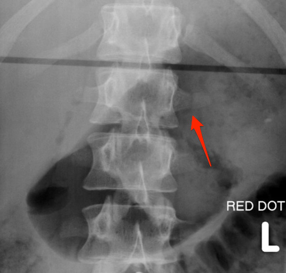


Thoracolumbar Spine X Rays
These areas look dark gray on the film A bone or a tumor is denser than soft tissue It does not let many Xrays to pass through and looks white on the Xray At a break (fracture) in a bone, the Xray beam passes through the broken area It is seen as a dark line in the white bone Xrays of the spine may be done to look at areas of the spineYour doctor may order a lumbar spine Xray to diagnose birth defects that affect the spine injury or fractures to the lower spine low back pain that's severe or lasts for more than four to eight weeks osteoarthritis, which is arthritis affecting the joints osteoporosis, which is a condition thatDue to xray beam divergence, it is necessary to include a projection of the thoracolumbar (TL) junction for a spinal radiographic survey that includes the thoracic and lumbar spine For the thoracolumbar junction lateral projection, position the patient in lateral recumbency ( Figure 3 )
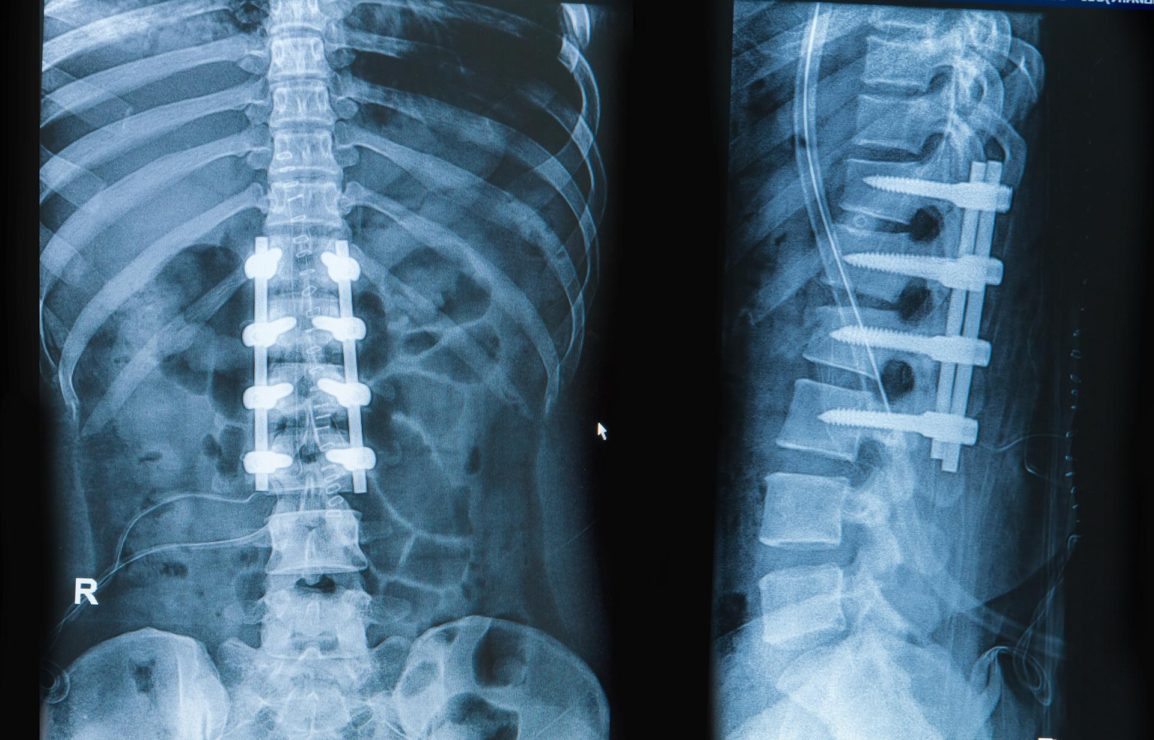


How Many Spinal Fusions Are Performed Each Year In The United States Idata Research


Xray Image Of Tl Spine Ap View Stock Photo Download Image Now Istock
A Thoracic Spine XRay may help diagnose (find) Thoracic spine Xrays can detect fractures in the thoracic vertebrae or dislocation of the joints between the vertebrae Xrays of the thoracic spine can also find the cause of tingling, numbness, or weakness in the arm or handThe XRay Lumber Spine test helps your doctor to check the bone structure of your lower back The lumbar spine comprises five vertebral bones The lower back of your pelvis has the sacrum, which is called the bone shield, located below the lumbar spine The lumbar spine also has the followingA thoracic spine Xray is an imaging test used to inspect any problems with the bones in the middle of your back An Xray uses small amounts of radiation to see the organs, tissues, and bones of
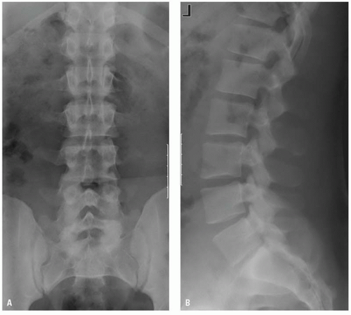


Imaging Thoracolumbar Spine Trauma Radiology Key


X Ray Image Of Human T L Spine Ap Lat Stock Photo Image By C V74
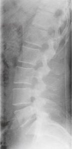
Lumbar Spine Xray Guideline Note For initial evaluation after trauma, routine 3 view (AP/Lateral/L5S1 Spot) is recommended unless requested by a spine surgeon Routine 3 views • AP • LATERAL (AP and LAT views should be centered on L3, and use proper collimation) • L5S1 SPOTA thoracic spine xray is an xray of the 12 chest (thoracic) bones (vertebrae) of the spine The vertebrae are separated by flat pads of cartilage called disks that provide a cushion between the bones How the Test is Performed The test is done in a hospital radiology department or in the health care provider's officeStanding AP and lateral Xrays are often ordered when back and/or leg pain doesn't go away The value of Xrays in diagnosing low back pain (LBP) has been questioned in the past In this study, researchers reviewed the use of flexionextension (F/E) Xrays for patients with LBP


The Radiology Assistant Thoracolumbar Injury


Thoracolumbar Spine Injuries Presentation And Treatment Bone And Spine
The lumbar spine series is comprised of two standard projections along with a range of additional projections depending on clinical indications The series is often utilized in the context of trauma, postoperative imaging and for chronic conditions such as ankylosing spondylosis Lumbar spine radiographs are one of the more commonly requested radiographic investigations of the spine, however, projectional radiography has limitations and further imaging such as MRI and CT should be consideredStanding AP and lateral Xrays are often ordered when back and/or leg pain doesn't go away The value of Xrays in diagnosing low back pain (LBP) has been questioned in the past In this study, researchers reviewed the use of flexionextension (F/E) Xrays for patients with LBPThoracoLumbar Spine Xray Guideline Routine 2 or 4 views • AP Thoracic spine • AP Lumbar spine • LATERAL Thoracic spine • LATERAL Lumbar spine • 2 VIEW is centered on junction of ThoracoLumbar spine • AP AND LATERAL VIEWS to include T8 thru L5 517 AMR


The Different Scoliosis Locations Types Of Spinal Curvatures Scolismart Blog


Radiology Of Burst Fractures Wheeless Textbook Of Orthopaedics
A 14" x 17" IR is used in the radiography of the adult Thoracic Spine (lengthwise/portrait) A 10" x 12" IR may be used for children/infants IR/Collimated Field Size A 14" x 17" IR is used in the radiography of the adult Thoracic Spine (lengthwise/portrait) A 10" x 12" IR may be used for children/infants 14Ejection from a motor vehicle or motorcycle;The thoracic spine xray is the imaging of the twelve bones extending from upper and middle back portion of our neck The thoracic spine joins the cervical spine and further connects with lumbar spine The Xray can spot for injuries in the thoracic spine In a human body a spine is made up of three subsections


A Case Of Paedatric Vertebral Compression Fracture Orthogate


Small Animal Spinal Radiography Series Thoracic Spine Radiography Today S Veterinary Practice
These areas look dark gray on the film A bone or a tumor is denser than soft tissue It does not let many Xrays to pass through and looks white on the Xray At a break (fracture) in a bone, the Xray beam passes through the broken area It is seen as a dark line in the white bone Xrays of the spine may be done to look at areas of the spinePhoto about Xray image of TL spine, AP view Showing a compression fracture at T12 Image of bone, joint, careLateral Lumbar spine is one of the basic projection and routinely taken in xray department Patient position is in lateral recumbent Film size use is 35 x 43 cm or 14 x 17 inches read more


The Thoracolumbar Spine
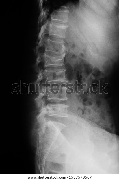


Thoracolumbar Spinetl Spine Xray Showing Spinal Stock Photo Edit Now
An xray of the lumbar spine reveals disc narrowing (desiccation), also known as degenerative disc disease, of both L 2,3 and L 3,4 Xray, Lumbar Spine Anteroposterior xray of the lumbar spine shows degenerative disc disease and mild scoliosis (curving of the spine) Desaturated color imageThis is positioning ONLY!Please remember to remove all obstructing items and, as always, practice proper radiation protection


Www Saintfrancis Org Wp Content Uploads Radiograph Spine Thoracic Vet Practice 13 Pdf
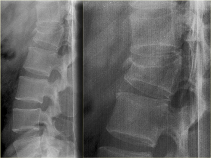


The Radiology Assistant Thoracolumbar Injury
A CT scan of the thoracic spine images the 12 vertebrae in the middle portion of the spine Your thoracic spine spans from the base of the neck to the middle of your back, running the length of your chest This exam is also called a Tspine CTA, An adequate lateral cervical spine xray, showing the entire cervical spine and its cephalad border (the skull base) and caudad border (T1) B, The image is inadequate because C1 and T1 are not seen fully Fractures or subluxation at these locations would be missedLumbar Spine Xray Guideline Note For initial evaluation after trauma, routine 3 view (AP/Lateral/L5S1 Spot) is recommended unless requested by a spine surgeon Routine 3 views • AP • LATERAL (AP and LAT views should be centered on L3, and use proper collimation) • L5S1 SPOT


Thoracolumbar Fracture Dislocation Spine Orthobullets


Ap Thoracic Spine Breathing Technique Wikiradiography
Dynamic Xrays of the Lumbar Spine How Useful Are They?Generally, an Xray procedure of the spine, neck, or back follows this process You will be asked to remove any clothing, jewelry, hairpins, eyeglasses, hearing aids, or other metal objects that may If you are asked to remove any clothing, you will be given a gown to wear You will be positioned


Xray Image Of Thoracolumbar Spine Stock Photo Download Image Now Istock


Ap Or Pa Projection In Lumbar Spine X Ray Radtechonduty


Ota Org Sites Files 18 08 S04 Thoracic and lumbar spine fractures and dislocations 28assessment 26 classification 29 Pdf


Q Tbn And9gctk3hklu0aynb8igztdob4olhkbmiz5akjdjyv Mp4wn Cqmham Usqp Cau


Q Tbn And9gcrxjf6zofwh6v50r1td A66v0grjlzzdidk6ve4juqbse3uky2x Usqp Cau


Xray Image Of Tl Spine Or Thoracolumbar Spine Ap And Lateral View For Diagnosis Back Pain Stock Photo Download Image Now Istock
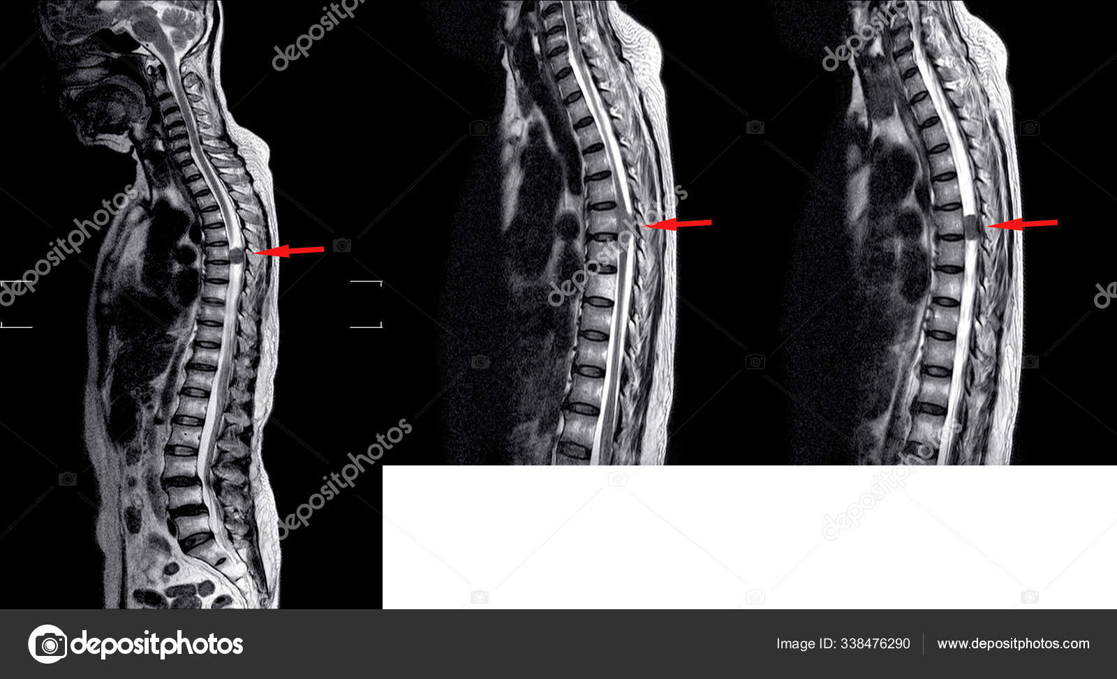


Mri Tl Spine Stock Photo Image By C Richmanphoto


X Ray T L Spine Stock Photo Picture And Royalty Free Image Image


Thoracolumbar Spine Lateral Canine X Ray Positioning Guide Imv Imaging Usa


The Thoracolumbar Spine


Thoracolumbar Spine X Rays
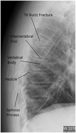


Case Study Management Of 58 Year Old Female With Burst Fracture Of T6 Vertebra


Traumatic Thoracolumbar Spine Injuries What The Spine Surgeon Wants To Know Radiographics


Surgical Correction Of Severe Adult Lumbar Scoliosis Major Curves 75 Retrospective Analysis With Minimum 2 Year Follow Up In Journal Of Neurosurgery Spine Volume 31 Issue 4 19
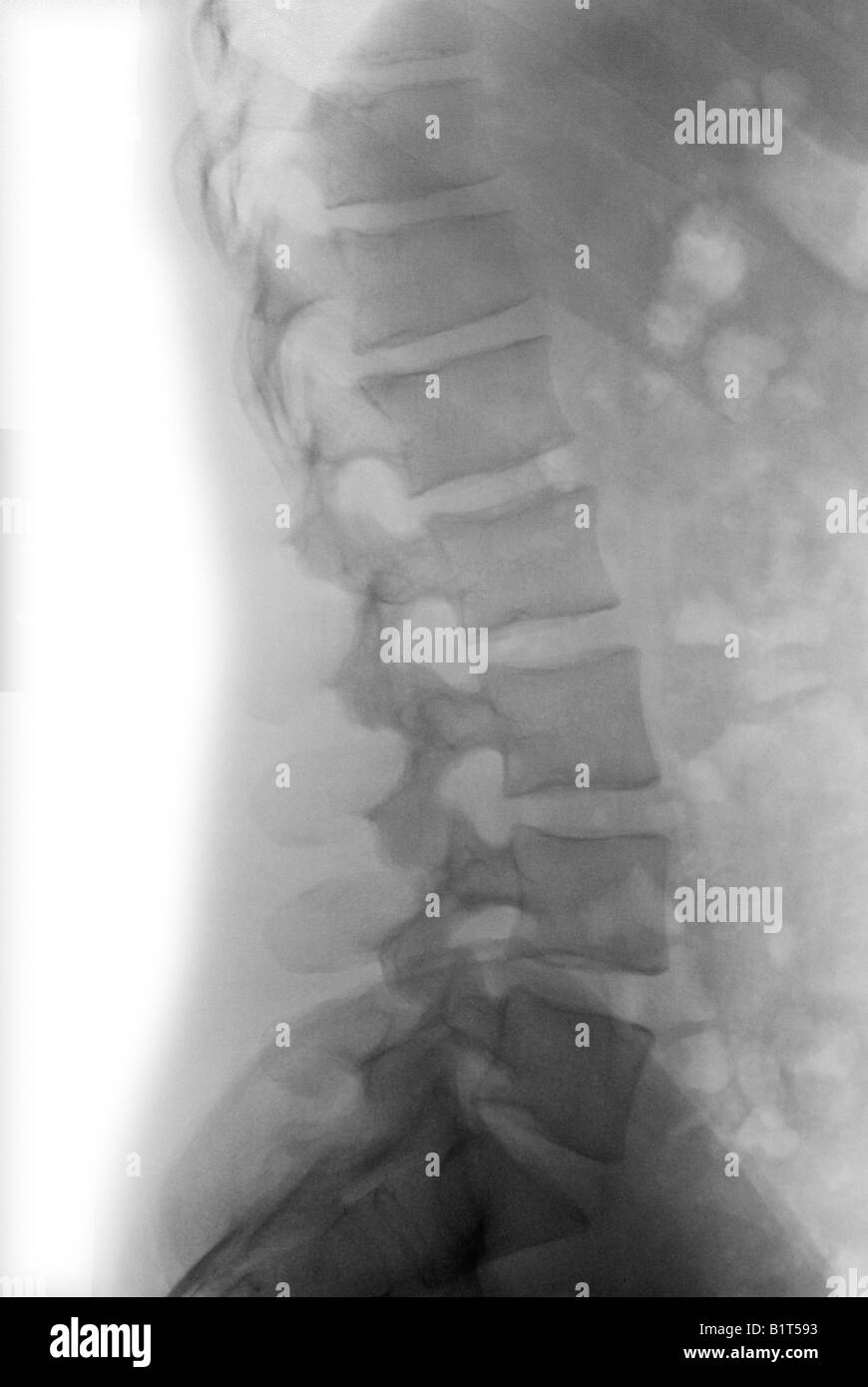


X Ray Normal Lumbar Spine High Resolution Stock Photography And Images Alamy


Miscellaneous


Thoraco Lumber Spine Ap Lat Anatomy And Physiology Part 17 Youtube
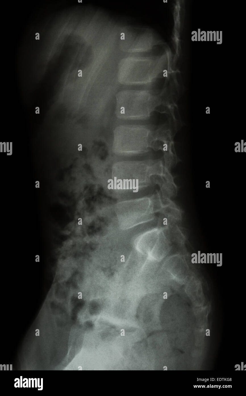


X Ray Normal Lumbar Spine High Resolution Stock Photography And Images Alamy


Http Www Oref Org Docs Default Source Default Document Library Sdsg Radiographic Measuremnt Manual Pdf Sfvrsn 2


Thoracolumbar Spine X Rays
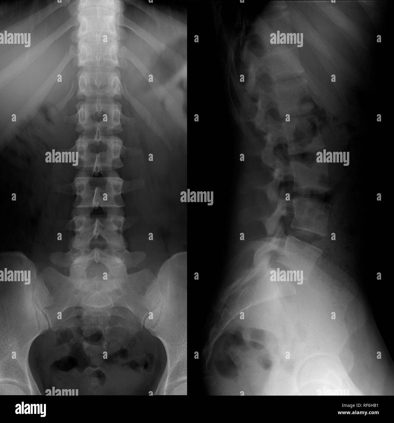


X Ray Normal Lumbar Spine High Resolution Stock Photography And Images Alamy


X Ray Positioning For Thoracic Spine Youtube


Thoracic Spine Ap View Radiology Reference Article Radiopaedia Org


Thoracolumbar Trauma With Delayed Presentation Kanna Rm Khurjekar K Indian Spine J


Lateral Lumbar Spine Radiography Wikiradiography


Thoracic Spine Series Radiology Reference Article Radiopaedia Org


Thoracolumbar Spine An Overview Sciencedirect Topics


Thoracolumbar Spine An Overview Sciencedirect Topics
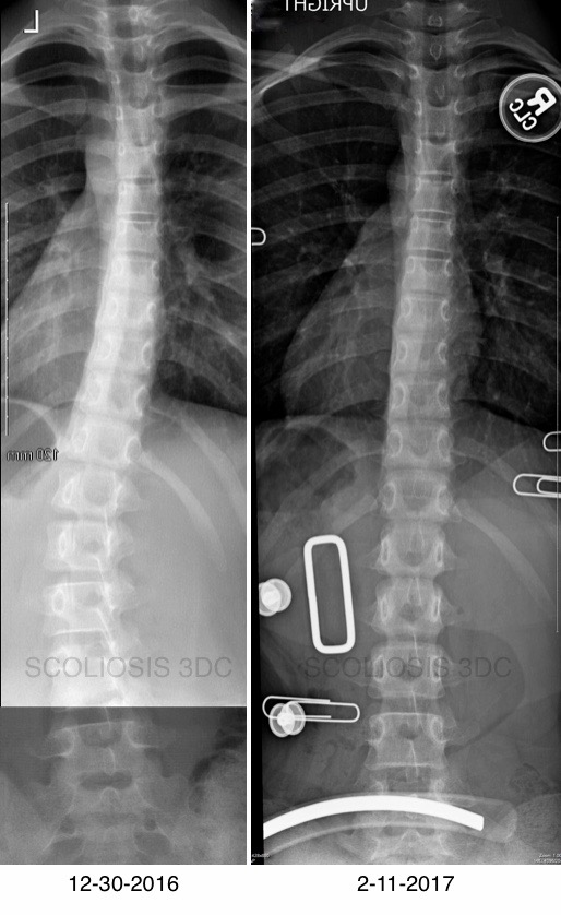


Thoracolumbar Scoliosis Scoliosis Of The Mid Spine


W1 Med Cmu Ac Th Ortho Images Education Dr Torpong Fractures dislocations of the spine Medical student v Pdf
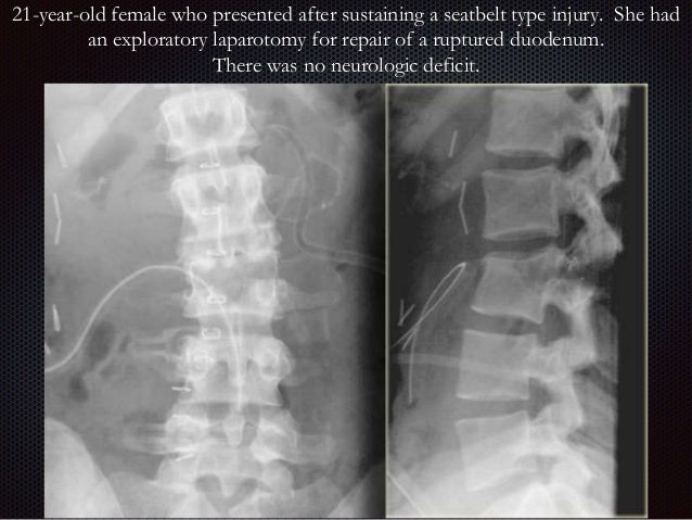


Imaging Of Thoracic Spine Trauma


Tl Spine X Ray Of Case 3 Tl Spine Lateral View Of Case 3 At Admission Download Scientific Diagram


Lateral T Spine X Ray Page 1 Line 17qq Com


Pediatric Thoracolumbar Spinal Injuries The Etiology And Clinical Spectrum Of An Uncommon Entity In Childhood Babu R A Arimappamagan A Pruthi N Bhat Di Arvinda H R Devi B I Somanna S Neurol India
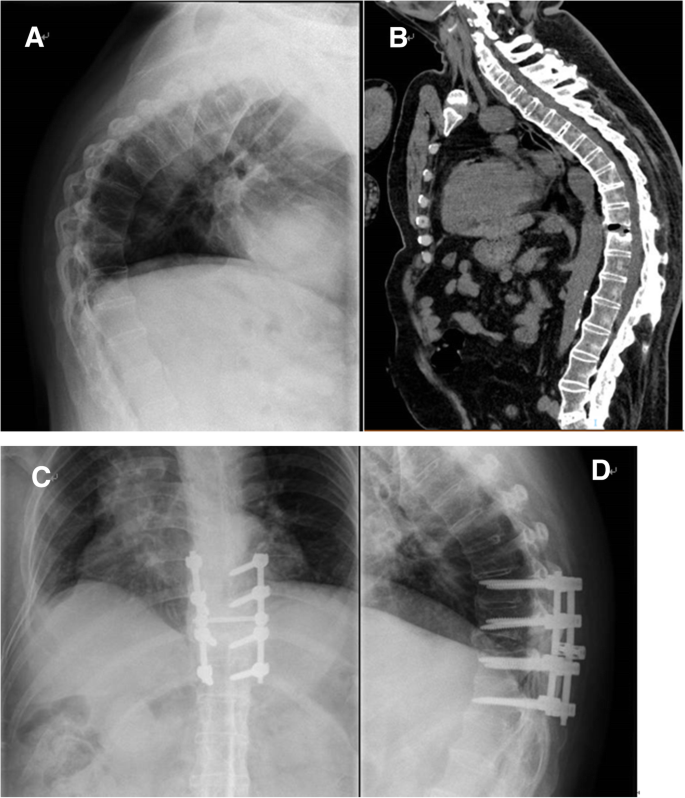


The Surgical Treatment Strategies For Thoracolumbar Spine Fractures With Ankylosing Spondylitis A Case Report Bmc Surgery Full Text


Lateral Lumbar Spine Radiography Wikiradiography


Www Saintfrancis Org Wp Content Uploads Radiograph Spine Thoracic Vet Practice 13 Pdf


Small Animal Spinal Radiography Series Thoracic Spine Radiography Today S Veterinary Practice
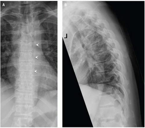


Imaging Thoracolumbar Spine Trauma Radiology Key


The Thoracolumbar Spine Radiology Key


Pediatric Thoracolumbar Spinal Injuries The Etiology And Clinical Spectrum Of An Uncommon Entity In Childhood Babu R A Arimappamagan A Pruthi N Bhat Di Arvinda H R Devi B I Somanna S Neurol India


Film X Ray T L Spine Thoracic Lumbar Spine Show Normal Human S Stock Photo Picture And Royalty Free Image Image
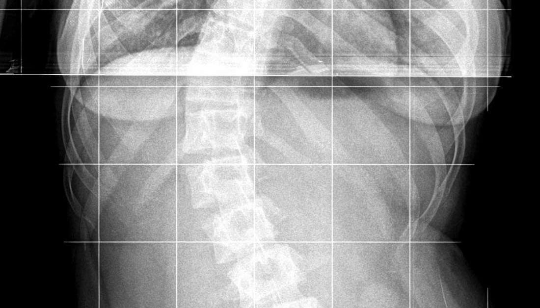


Dextroscoliosis Types Exercises And Treatment


Thoracolumbar Fracture Dislocation Spine Orthobullets


X Ray T L Spine Stock Photo Picture And Royalty Free Image Image
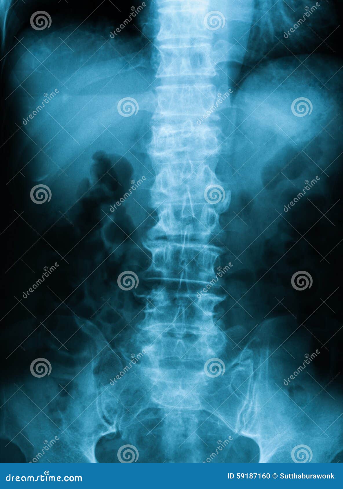


X Ray Image Of T L Spine Apview Stock Photo Image Of Bone Joint
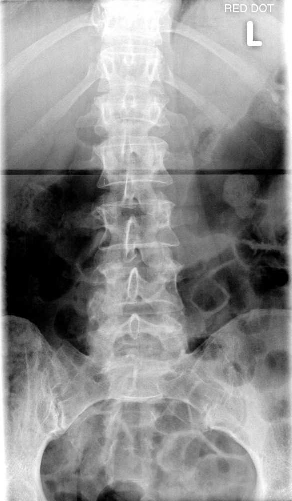


The Thoracolumbar Spine
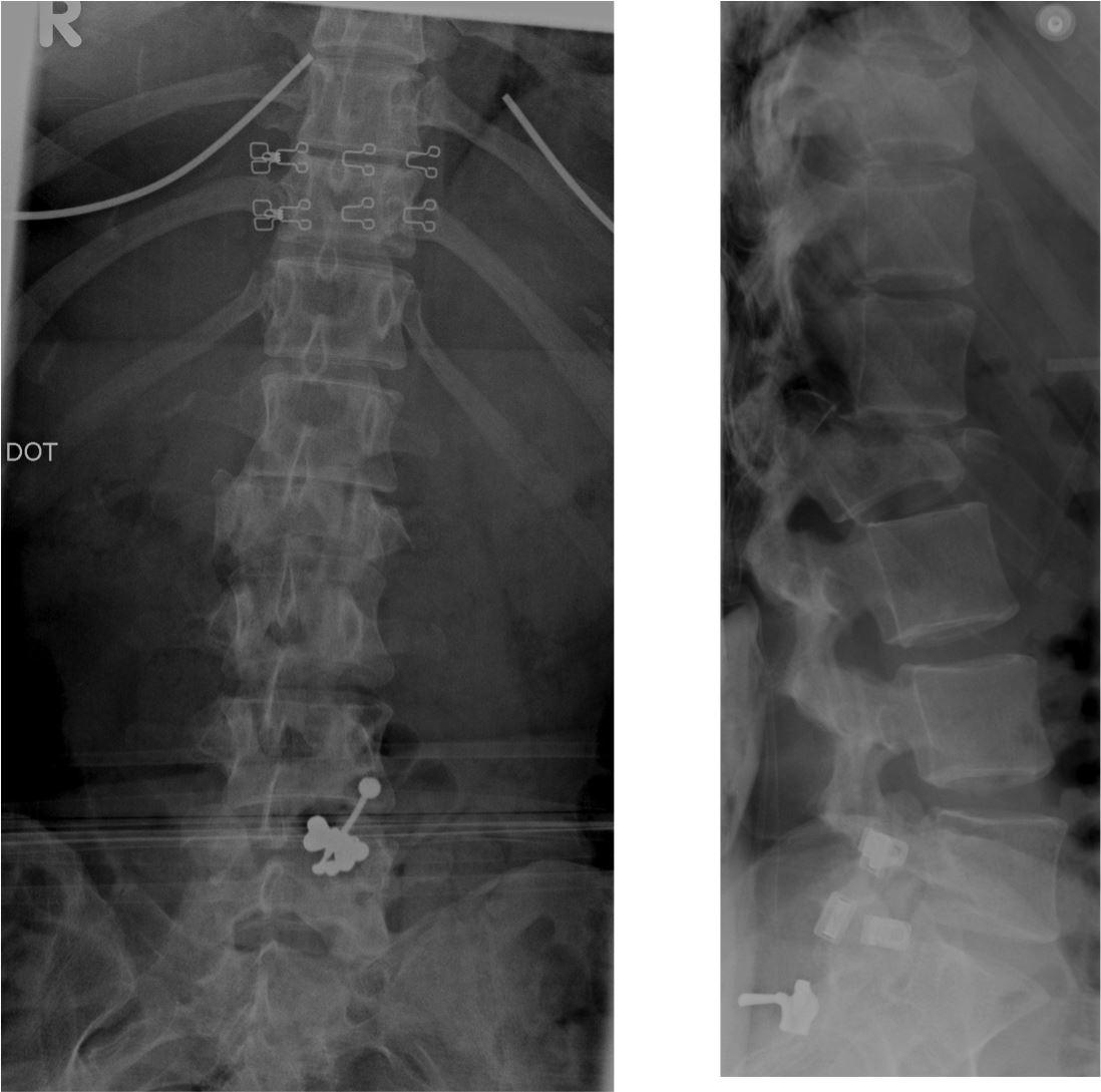


The Thoracolumbar Spine


Xray Image Of Tl Spine Lateral View Stock Photo Download Image Now Istock


Q Tbn And9gcs1iefur4rbtrf3xl8whgxvoft Him9uue0ntcrrx X1dzvd3jh Usqp Cau
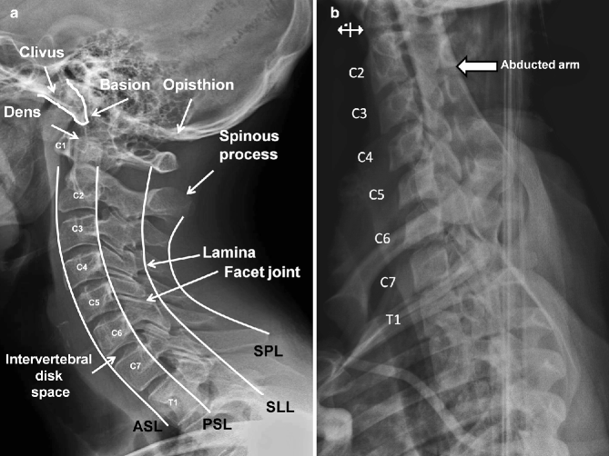


Radiologic Imaging Of The Spine Springerlink


Thoraco Lumbar Fracture The Bone School


Ce4rt Radiographic Positioning Of The Lumbar Spine For X Ray Techs


The Thoracolumbar Spine


Soft Blurry Xray Tl Spinethoracospinepost Fixed Stock Photo Edit Now


Oregon Providence Org Media Images Modules News Ptkattachments News Xr ordering guide 13 Pdf


Thoracolumbar Junction Lateral Spine Dislocation Sciencedirect


Anteroposterior And Lateral X Rays Of The T L Spine Show A T12 Download Scientific Diagram


X Ray Image Of T L Spine Or Thoracolumbar Spine Lateral View For Diagnosis Back Pain Buy This Stock Photo And Explore Similar Images At Adobe Stock Adobe Stock


The Thoracolumbar Spine


Scoliosis Musculoskeletal Key


A Novel Radiographic Classification Of Severe Spinal Curvatures Exceeding 100 The Omega W Gamma G And Alpha A Deformities Sogacot


Thoracolumbar Fracture Dislocation Spine Orthobullets


The Surgical Treatment Strategies For Thoracolumbar Spine Fractures With Ankylosing Spondylitis A Case Report Bmc Surgery Full Text


The Thoracolumbar Spine


Thoracolumbar Fractures Changing Perspectives International Journal Of Spine


A Case Of Paedatric Vertebral Compression Fracture Orthogate


Normal Thoracic Spine Radiology Case Radiopaedia Org


Flexion Distraction Injuries Of The Thoracolumbar Spine Open Fusion Versus Percutaneous Pedicle Screw Fixation In Neurosurgical Focus Volume 35 Issue 2 13
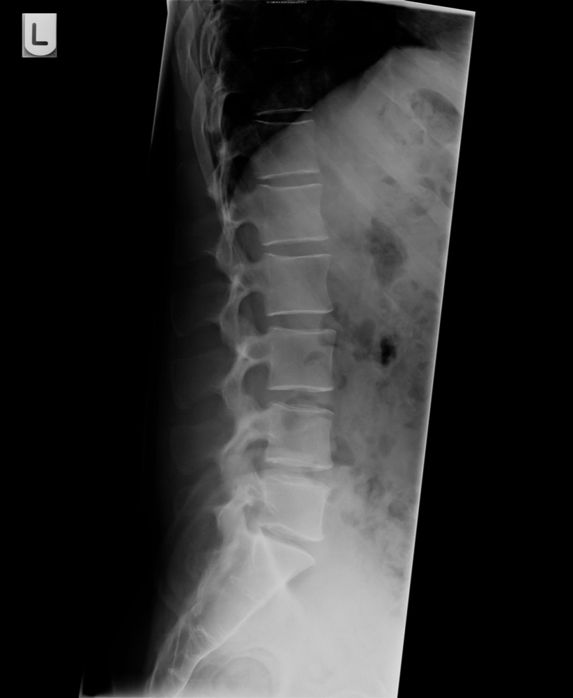


The Thoracolumbar Spine


Colorful X Ray T L Spine Thoracic Lumb Ar Spine Canstock


A Lumbar Spine X Ray In The Hyperflexion Position The Vertebrae From Download Scientific Diagram


Dextroscoliosis Types Exercises And Treatment


Http Pdf Posterng Netkey At Download Index Php Module Get Pdf By Id Poster Id 1149


The Thoracolumbar Spine Radiology Key


Thoracic Spine Lateral Canine Veterinary X Ray Positioning Guide Imv Imaging


コメント
コメントを投稿